

Biosensor measures separate molecules in blood
Engineers at Eindhoven University of Technology have developed a biosensor that can register individual molecules in blood. This allows doctors and laboratory staff to measure the concentration of proteins, hormones, etc., very accurately.
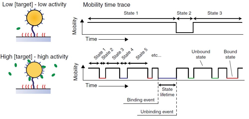
The biomarker technology developed by Eindhoven University of Technology works by means of mutations in the mobility of the biosensor. This relates to the combination of a particle smaller than a micrometre which has certain biomarkers, and a substrate also containing biomarkers. That particle is attached to the substrate by means of a flexible DNA string.
'If the blood contains a target molecule to which the biosensor is sensitive, it will become attached to both the particle and the substrate,' explains Professor Menno Prins of the Biomedical Engineering Department of Eindhoven University of Technology.
Selective
This approach offers two benefits. 'We're very selective, as the target molecule must become attached to two different biomarkers.'

This method also makes the attachment of the target molecule discernible. 'Due to the double attachment, the mobility of the biomarker in the blood changes, and that is easily detectable.' This is due to the size of the particle (less than a micrometre) versus that of the target molecule (one-hundredth of a micrometre at most). With only a single attachment, there would be very little difference in the particle movement, whereas its attraction to the substrate results in a discernible change.
As this attachment concerns a dynamic balance, the target molecule will become unattached again. 'It becomes a digital on and off signal as it were, and the degree at which it changes relates to the concentration of the target molecules in the blood.'
Light source, lens and algorithm

It's relatively simple to observe the change in movements. The large particle containing the biomarkers has been created to clearly light up under a microscope. And so the change in movement can be clearly followed through a microscope. 'All you need is a light source, a camera and an algorithm that indicates whether the particle is moving freely or is attached,' explains Prins. Using these results, doctors can determine the concentration of the target substance in the blood.
This monitoring process can take place continually, which is another important benefit of our method.' It is conducted via a catheter, for example: in hospitals, there are many treatments which require one to be used, and the detector can then be attached directly to the catheter in place.
The biomarker technology is already so advanced that it is available for all kinds of target molecules.
Now that the Eindhoven engineers have proven that the method works, they are looking to design a more practical version of the detector.
If you found this article interesting, subscribe for free to our weekly newsletter!


Reacties