

Proof at last: Van Leeuwenhoek ground his microscope lenses
Researchers from Delft University of Technology and Rijksmuseum Boerhaave have solved a centuries-old puzzle regarding the microscopes of Antoni van Leeuwenhoek. He used thin lenses that he ground himself.
Given the unparalleled quality of the microscope images that Van Leeuwenhoek produced, up till now this had always been considered practically impossible. According to conventional wisdom, it was simply a step too far to believe that small lenses could be so perfectly ground by hand. The solution to this mystery is down to a new research method: the use of a neutron beam originating from Delft University of Technology's research reactor.
Advanced method
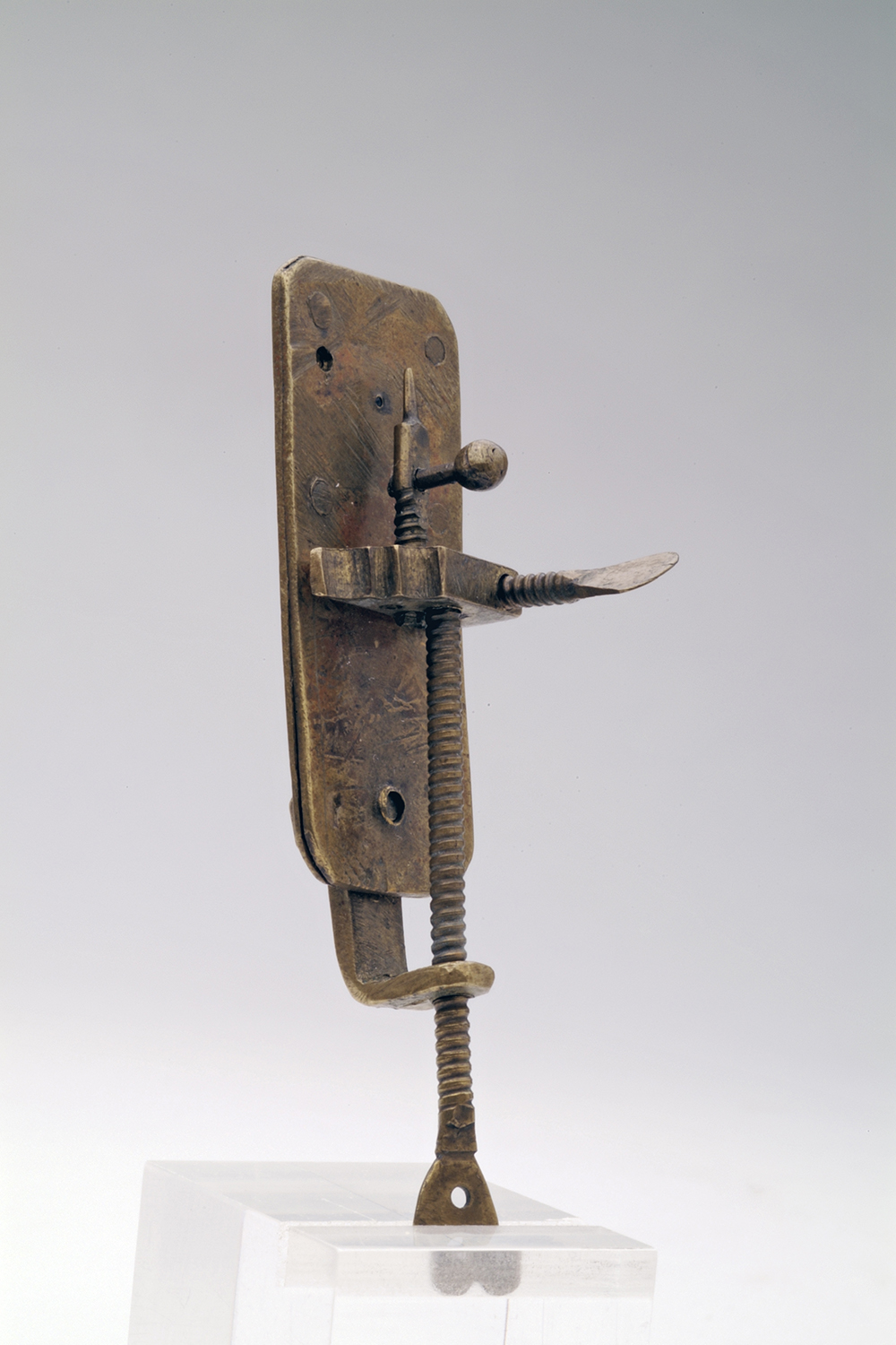
The microscopes made by Antoni van Leeuwenhoek (1632 - 1723) comprised a single lens with a ‘skewer’ on which he would impale the object to be studied. Whereas the devices of his contemporaries managed to reach a magnification of around thirty times, Van Leeuwenhoek made microscopes up to ten times that power. Was his claim true that he had invented an advanced glass-blowing method, something he revealed in 1711 to a group of German nobility in a rare moment of candour? Or was the quality of the lens simply down to meticulous grinding?
The eleven ‘Leeuwenhoeks’ that have survived – four of which are found in the Boerhaave collection – carry the secret within them. ‘Van Leeuwenhoek clamped his lenses between two metal plates that he sealed using rivets,’ says Tiemen Cocquyt, museum curator and participant in the study. ‘Given their rarity and enormous historical value, disassembly is not an option. Apart from a half-millimetre hole, the lenses are inaccessible. Over ninety percent is simply not visible. And that's been the case for 350 years already.’
Particles without charge
The puzzle of the Leeuwenhoek lens has been solved using new technology. This makes it possible to build up a picture of the inside of a microscope without having to break it open. This non-invasive method of imaging is known as neutron tomography. The Delft Reactor Institute has a new instrument that works on the basis of this technology.
‘Tomography involves rotating an object in a neutron beam in front of a camera while taking pictures,’ explains Delft University of Technology researcher Lambert van Eijck. ‘Neutron particles do not have an electrical charge, and unlike X-rays for example, will penetrate metal. After rotating the object 180 degrees, you can then use the 2D images produced to construct 3D images of the object on a computer.’
Skilled grinder
The image that Van Eijck has made of one of the Boerhaave microscopes using this method leaves no more room for doubt: Leeuwenhoek lenses were not blown, but ground. ‘It turns out that there really was no exotic production method after all, but Van Leeuwenhoek was simply remarkably skilled in grinding his minuscule lenses,’ says Cocquyt.
One question that the researchers would still like to answer is whether the lens is made of a special type of glass. ‘We can investigate this using gamma spectroscopy,’ explained Van Eijck. ‘Neutron tomography makes an object temporarily radioactive. The way in which the radioactivity decays reveals which elements the object contains.’
If you found this article interesting, subscribe for free to our weekly newsletter!
Opening photo: The neutron image of the Leeuwenhoek lens.


Reacties