

The art of science
In an exhibition called True Beauty – Where science and art meet, the walls of the Stedelijk Museum Breda are adorned with scientific images as if they were true works of art. 'Some researchers give their work an almost artistic allure that is more at home in a museum than a laboratory.' The exhibition just opened and will run through Sunday 19 August.
‘Scientists are increasingly able to visualise all kinds of things,’ explains Dingeman Kuilman, Managing Director of the Stedelijk Museum Breda. 'Whether it's the size of an atom or has the dimensions of the atmosphere, they can visualise what the naked eye could never see.'
Kuilman talks as he walks around the exhibition gallery of True Beauty, where work is still under way on the exhibition a few days before the opening. This is the first large exhibition staged at this museum, which opened its doors for the first time in the summer of 2017. Kuilman searched the databases of research institutes all over the world to find the True Beauty material. Animatedly, he explains all about the works on show and precisely why they were chosen.
‘Many scientific images are no longer photos, but actually the visualisation of data. Researchers process information to create images of the invisible reality. They combine objective measurements with the power of imagination, and actually make artistic decisions in doing so. Electron microscopy produces a type of RAW file for example, containing raw and unprocessed data. The decisions on colour, lighting and cut then determine the final quality of the image.'
Brain connections
Kuilman pauses at an image of a so-called 'connectonome' that shows the route taken by the various nerve pathways in the brain and how they interconnect. This started with an MRI scan of the brain. The raw data comprised slices – cross sections – of the brain. These were then coupled to each other and processed in a number of steps, resulting in this object.
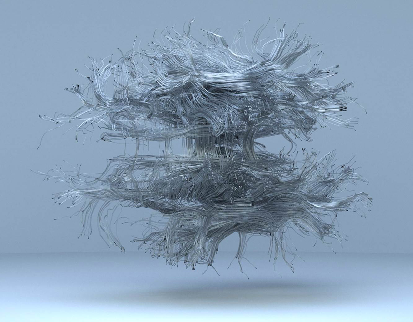
'The researcher, David Liewald of the University of Edinburgh, chose to suspend the connectonome in space. He's even given it a shadow, which makes it an object with a surrealist look. It's fascinating how he's semi-consciously chosen this presentation,’ according to Kuilman.
Choice of colours
Kuilman then points out a canvas which is reminiscent of a Van Gogh, but is actually a microscopic image of a great diving beetle. We can see the male's suckers on his front legs, clinging to the female's wing case during mating. The choice of colours in the microscopic image is reminiscent of an expressionist painting.
'This is mainly an aesthetic choice. There are also scientific images which require the addition of colour and texture in order to render items visible. And so the researcher is forced to give their own interpretation.'
To illustrate this, he mentions the image of a minute blood vessel which is found behind our eye. Researcher Peter Maloca of Basel University in Switzerland visualised it using optical coherence tomography. It is a digital reconstruction of more than 250 microscopic cross sections which have been mounted in succession. The colour and texture were then added artificially.


Kuilman has hung the eye socket next to an image derived from the science fiction film Fantastic Voyage dating from 1966. In the film, a submarine and five crew members were reduced to the approximate size of a microbe before being injected into the body of a leading scientist. Their task is to remove a blood clot from his brain in order to save his life. ‘The two images emanate the same atmosphere. You'd almost think that Maloca had based it on Fantastic Voyage.’
Print room
There is but a limited explanation to accompany the images in the exhibition gallery, with only a few key words given. ‘They're search terms as it were,’ says Kuilman. The more detailed description is given in a guide provided for visitors. This was a conscious choice, Kuilman explains. 'First and foremost, this is an exhibition of pictures in an art museum, rather than a scientific exhibition.’
Careful thought was therefore also given to how the scientific images should be presented. 'We've consciously displayed them without frames or passe-partouts, as that would give the images a status which they themselves do not have. Neither were they intended for that. We also chose not to make them too large, but rather to present them in the style of a print room.'
The boundaries between art and science are blurring
The visualisations by international top researchers hang amiably shoulder to shoulder with works by artists inspired by science. Pride of place is for the Breda-born artist Hubert Leyendeckers, whose work is reminiscent of galaxies.
'The boundaries between art and science are blurring,' Kuilman concludes. 'This exhibition is proof of the new grey area.'
The exhibition True Beauty – Where science and art meet is on from Saturday 3 March through Sunday 19 August 2018 in the Stedelijk Museum Breda.
If you found this article interesting, subscribe for free to our weekly newsletter!
Opening image: Great diving beetle, Spike Walker


Reacties