
Microscope makes 3d videos of living cells
For many years, biologists were unable to effectively study living cells in action, but that is now changing thanks to a pioneering development by American physicists and engineers. They combined three microscopy techniques in one device that makes crystal-clear images of cells at work.
Until now, scientists were able to study living cells in their natural environment only by illuminating them with a very bright light and then viewing them through a microscope. But this bright light causes cells stress and they no longer behave as they would naturally.
A team of researchers headed by the Howard Hughes Medical Institute has now discovered a solution to this problem. They built the prototype of a microscope that actually combines three different microscopy techniques. This device makes crystal-clear and colourful images of all kinds of cells and tissues, without needing such an enormous amount of light.
The new microscope provides fascinating images. Here an immune cell (orange) can be seen as it moves through the inner ear of a zebra fish, picking up sugar particles (blue) as it goes.
Compensation of distortions
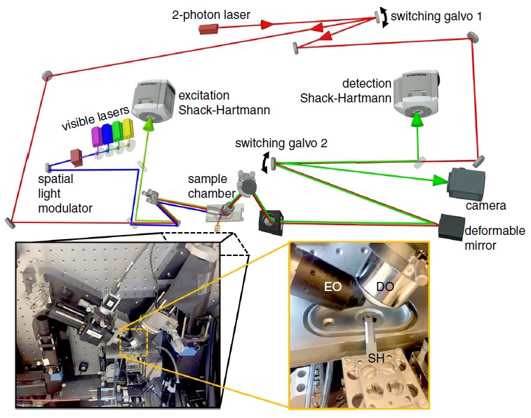
Firstly, the new microscope (see illustration below) comprises an adaptive optics system inspired by telescopes. These optical elements compensate distortions in the image that are for instance caused by the light passing through various tissues.
The second important component of the microscope is a clever way of illumination referred to as lattice light sheet microscopy. In this method, a sheet of light is repeatedly moved quickly through the specimen. A computer program then compiles the resulting series of 2D images to form a three-dimensional image.
Finally, the microscope comprises a laser bundle that determines the optical deformations (aberrations) at every point on the specimen, so that these can be compensated by the software. These deformations differ for every type of tissue. The laser thus serves to correct the deviations that occur using the two microscopy techniques described.
Optical panel
The physicists and engineers have merged these three techniques into a sophisticated test configuration that currently still stands on a 3-m-long optical panel. With such a format, the microscope is not yet very suitable for use in laboratories. But the inventors are already working on miniaturising the whole thing and on making it more user-friendly and more affordable. ‘All the technical demonstrations and publications don’t stand for much. A microscope is only really good when a lot of people use it,’ says project manager Eric Betzig from the Howard Hughes Medical Institute.
The first real microscope with the techniques described on board will be installed at the Advanced Imaging Center of the Janelia Research Center at the Howard Hughes Medical Institute in Virginia, USA. Scientists from all over the world can then apply to come and study their specimens at the Center. The Americans also want to develop a microscope with their ideas that will be commercially available.
In the meantime, admire the images of a human breast cancer cell as it moves through a piece of tissue of a zebra fish:
Take a look also at this collection of videos made by the inventors of the new microscope:
Opening image: Image of the spine of a young zebra fish. New neurons that are produced light up in different colours. All photos: T. Liu et al., Science, 2018.
If you found this article interesting, subscribe for free to our weekly newsletter!
Meer artikelen

Een AI-fabriek in Groningen

Gezondheid meten via zweetdruppels


Nieuwste artikelen
Een AI-fabriek in Groningen


