
Organs projected onto the body
A team of physicians and programmers in Canada have developed augmented-reality (AR) software that projects the body's internal organs onto the skin. The 3D images are calculated using different CT and MRI scans. The new AR technique makes the inside of a patient's body clearly visible and can be used for educational purposes, surgical planning and consultations between doctors.
Wouldn't it be great to be able to look right through your skin and see your organs? We already have anatomical mannequins of course, which can be opened up and the organs removed one by one. And two-dimensional medical scans (ultrasound, CT scan, MRI scan) are standard tools in hospitals these days. But projecting the inside of a living person onto their skin? That was just fantasy, until now.
Doctors, researchers and programmers from the University of Alberta in Canada have developed software, called ProjectDR, which uses CT and MRI scans to calculate colourful 3D images and then projects them onto the patient's body. The images even move together with the body, if it moves.
Different colours
 This is a new way of looking inside the body without having to open it up using a surgical blade. And though it seems to be a fancy way of pimping medical scans, it is far more than just that. Because the various types of tissue and organs are assigned different colours, it is possible to switch organs ‘on’ or ‘off’ in this project. Just want to look at the lungs? That's easy. Or just the blood vessels? No problem, with the simple press of a button.
Education and operation planning
In the first place, that creates added value for training prospective doctors. After all, pictures of people's flesh and blood provide far more information than anatomic mannequins. But planning operations could also benefit from the extra information provided by such a projection of someone's unique internal set-up – because no two people are alike.
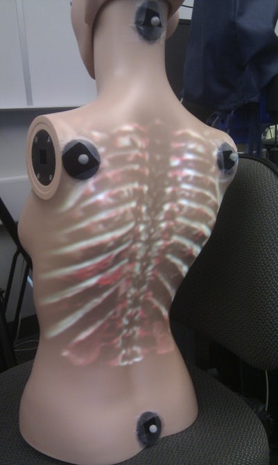
Markers
To ensure that the images move with the patient, the developers have used motion tracking. A number of markers are placed on the body (to the right on the photo) that reflect infra-red light. If the patient's posture changes, an infra-red camera observes that directly and adjusts the projected images accordingly.
The people involved stressed that the project is still in the research phase. And that can be seen in the video below: although the projection does adjust with the movement, there is clearly somewhat of a delay. This is one of the matters the researchers are currently working on: faster feedback. They will also be using depth sensors to measure body movement even more accurately.
Tests in the operating theatre
The ProjectDR system will then be tested in a hospital setting. In the operating theatre, surgeons and nursing staff will study the pros and cons of body projections. ‘But we are also doing trials with chiropractors and physiotherapists to determine what the added value could be for their practices’, says Greg Kawchuk, Professor of Recovery Therapy at the University of Alberta.
If you found this article interesting, subscribe for free to our weekly newsletter!
Image material from University of Alberta
Meer artikelen

Een AI-fabriek in Groningen

Gezondheid meten via zweetdruppels


Nieuwste artikelen
Een AI-fabriek in Groningen


